

Equipment
Automatic Special Stainer - BenchMark Special Stains
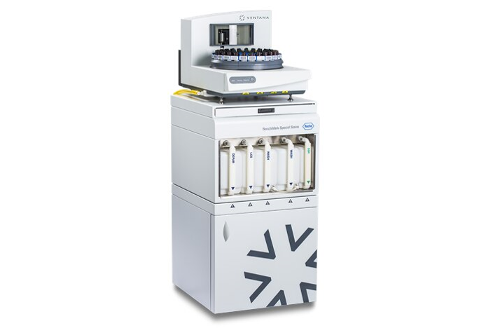
| Model | BenchMark Special Stains |
|---|---|
| Manufacturer | Roche |
| Year Introduced | 2020 |
| Usage Type | Analysis Request (paid) |
| Features |
|
| Usage | Available through analysis service request |
| Location | Medical Research Innovation Center (CMI), Room B2305 |
Automatic Stainer and Coverslipper - HISTOCORE CHROMAX ST CVWS
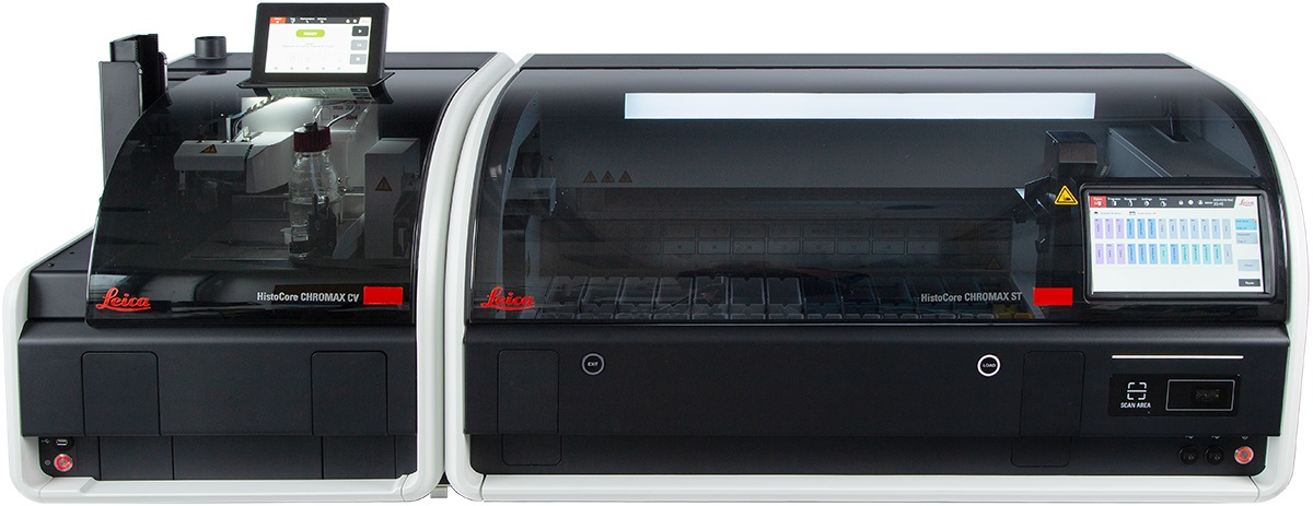
| Model | HISTOCORE CHROMAX ST CVWS |
|---|---|
| Manufacturer | Leica Biosystems |
| Year Introduced | 2025 |
| Usage Type | Analysis Request (paid) |
| Features | A staining instrument used for observing tissue slide specimens; a basic stainer that automatically performs H&E staining, deparaffinization, dehydration, and mounting during slide preparation |
| Usage | Available through analysis service request |
| Location | Medical Research Innovation Center (CMI), Room B2305 |
Automatic Rotary Microtome - RM2255
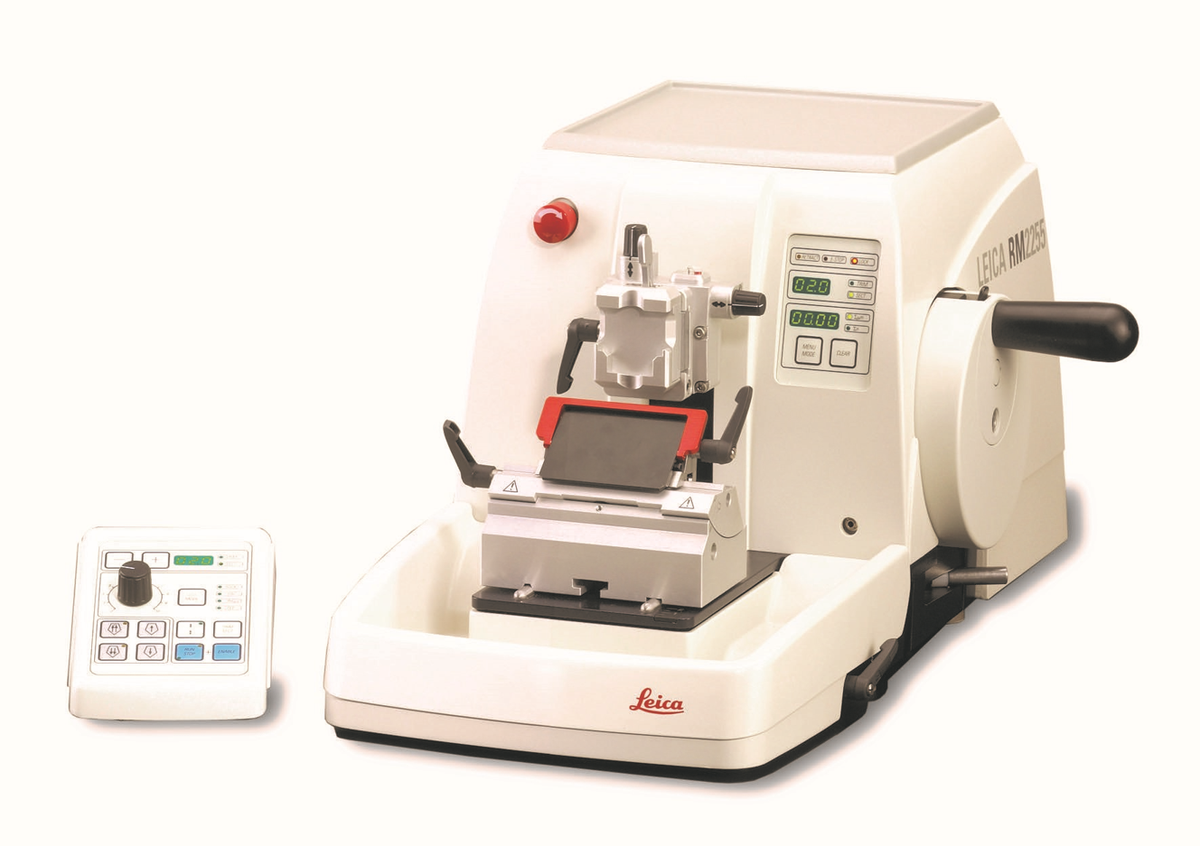
| Model | RM2255 |
|---|---|
| Manufacturer | Leica Biosystems |
| Year Introduced | 2016 |
| Usage Type | Analysis Request (paid), Personal Use (paid) |
| Features | A microtome is a core instrument that, after preparing a tissue specimen into a paraffin block, sections it into extremely thin slices on the micrometer (µm) scale so that it can be observed under a microscope |
| Usage | Refer to the reservation site for usage precautions and the no-show policy |
| Location | Medical Research Innovation Center (CMI), Room B2305 |
Tissue Processor - HistoCore PEGASUS
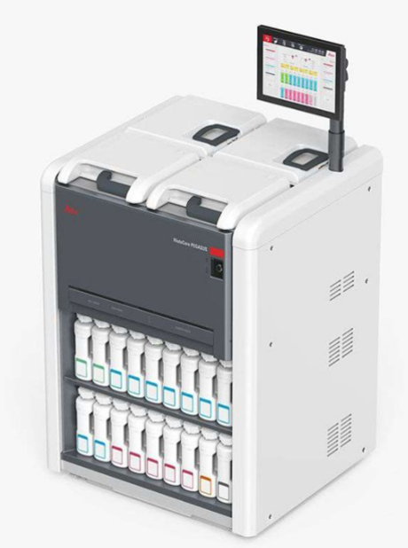
| Model | HistoCore PEGASUS |
|---|---|
| Manufacturer | Leica Biosystems |
| Year Introduced | 2024 |
| Usage Type | Analysis Request (paid) |
| Features | An instrument that automatically performs the dehydration–clearing–infiltration steps required to process animal or human tissue, from fixation to a state fully infiltrated with appropriate histological paraffin, so that it can be embedded for sectioning on a microtome |
| Usage | Available through analysis service request |
| Location | Medical Research Innovation Center (CMI), Room B2305 |
Automatic Immunohistochemistry Stainer - Ultra DISCOVERY XT
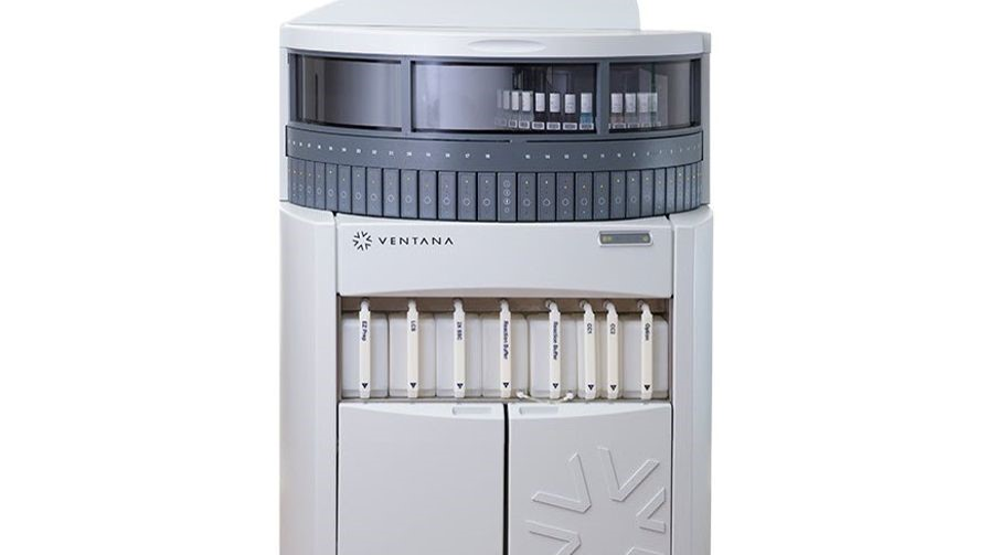
| Model | Ultra DISCOVERY XT |
|---|---|
| Manufacturer | Ventana |
| Year Introduced | 2019 |
| Usage Type | Analysis Request (paid) |
| Features |
|
| Usage | Available through analysis service request |
| Location | Medical Research Innovation Center (CMI), Room B2305 |
Cryostat Microtome - CM1950
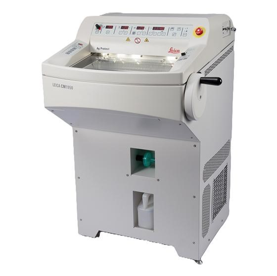
| Model | CM1950 |
|---|---|
| Manufacturer | Leica |
| Year Introduced | 2015 |
| Usage Type | Analysis Request (paid), Personal Use (paid) |
| Features |
|
| Usage | Refer to the reservation site for usage precautions and the no-show policy |
| Location | Medical Research Innovation Center (CMI), Room B2305 |
Digital Slide Scanner - Aperio GT450DX
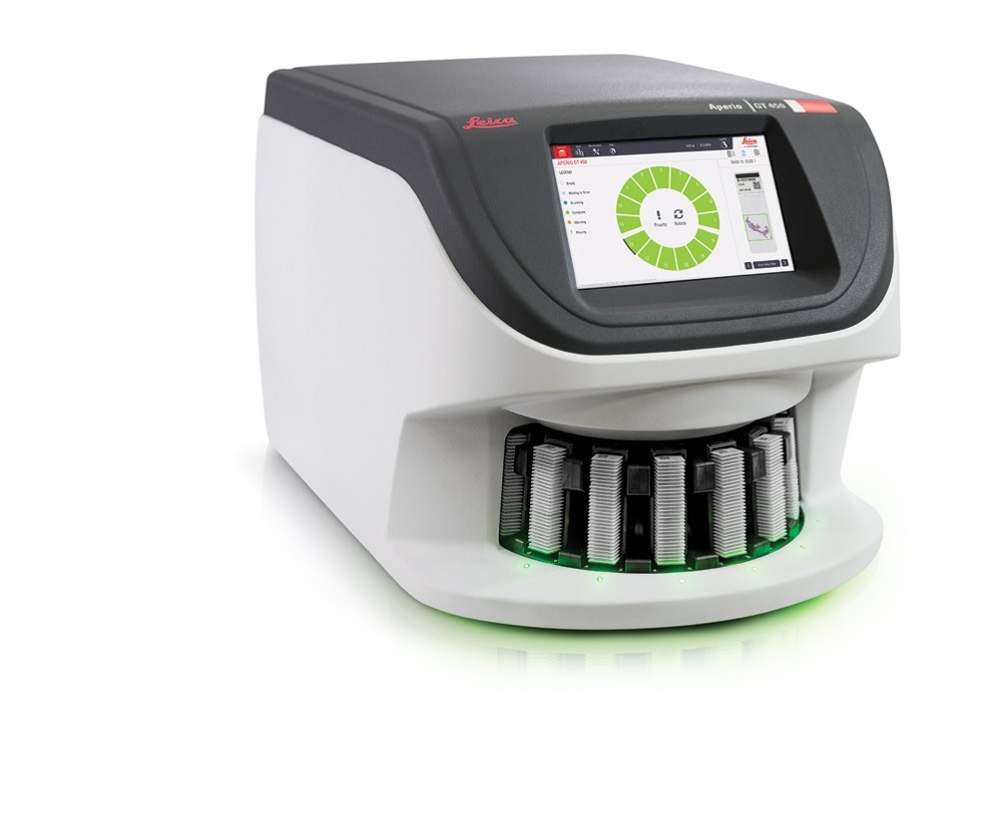
| Model | Aperio GT450DX |
|---|---|
| Manufacturer | Leica |
| Year Introduced | 2024 |
| Usage Type | Analysis Request (paid) |
| Features |
|
| Usage | Available through analysis service request |
| Location | Medical Research Innovation Center (CMI), Room B2305 |