

About
Overview
An advanced Cell Imaging & Metabolism research platform that quantitatively analyzes the physiological changes, functions, and energy metabolism of cells
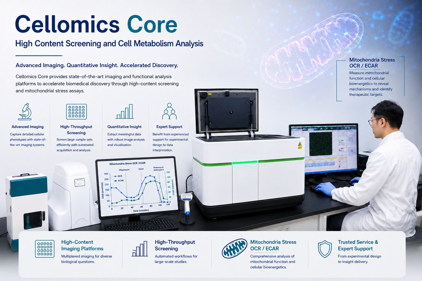
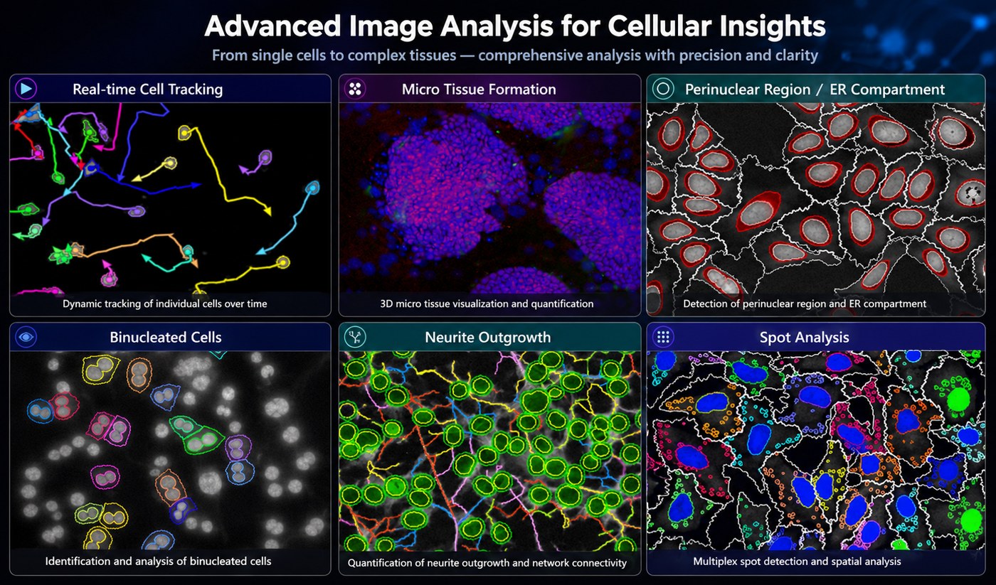
Cellomics is an “integrated bioimaging analysis field” that quantitatively analyzes cell morphology, function, and signal transduction using multi-well plate imaging technology based on High-Content Screening (HCS) / High-Content Analysis (HCA). By automatically acquiring cell images and interpreting them through objective data analysis and visualization, biological changes occurring at the cellular level can be quantitatively understood.
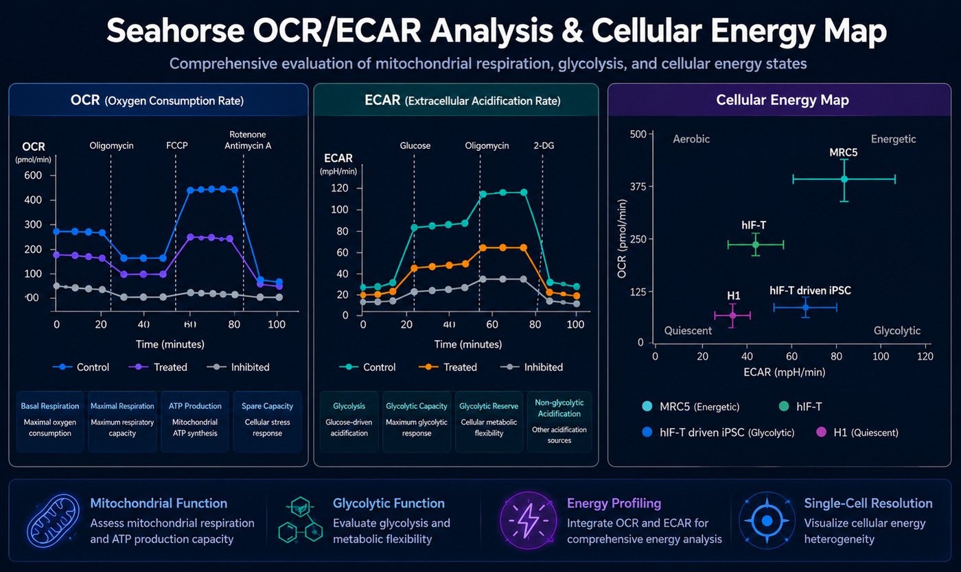
The Cytomics Laboratory of the Translational Medicine Research Institute at Seoul National University Hospital performs not only advanced HCS-based image analysis technology but also mitochondrial stress and cell metabolism research through Seahorse XF-based OCR/ECAR analysis, cell-to-cell signal transduction research through extracellular vesicle (EV) analysis, and multi-well plate-based cell function analysis, providing analytical techniques and academic support related to research on cell function, energy metabolism, and cell-to-cell communication.
In addition, it provides experimental design consulting and data interpretation support tailored to each researcher's experimental objectives, thereby enhancing the efficiency and completeness of research.